More, with Orca™

AI Powered, Seamlessly Integrated

AI model hub to get you started

AI model hub to get you started

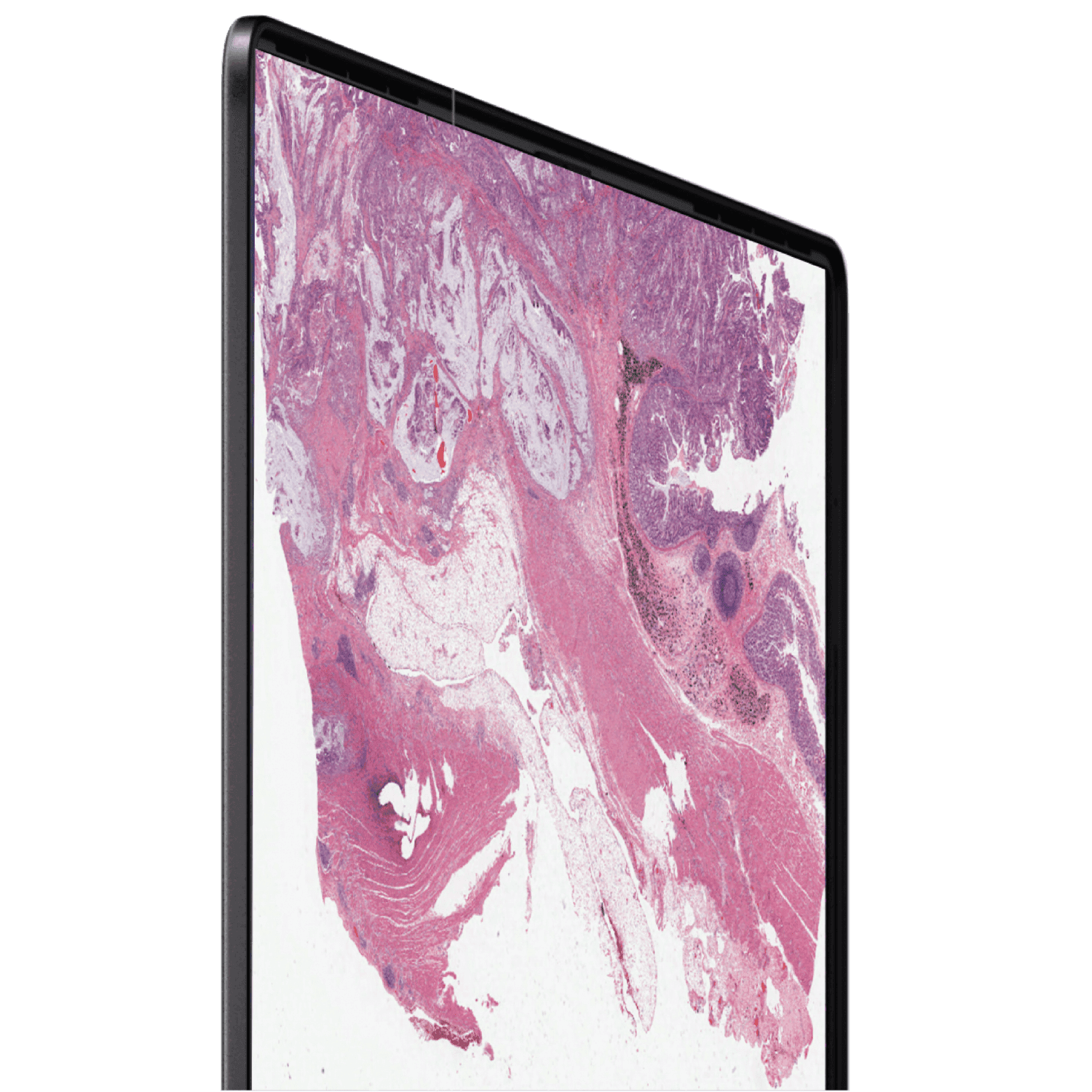
Achieve unprecedented scale in whole-slide image analysis with our ready-to-deploy AI models. ORCA: Jumpstart your research with our diverse collection of pretrained AI models for cancer and cell classification. Our solutions offer the functionality, scalability, and user-friendliness needed to seamlessly integrate AI into your research and daily workflow.

The use of models in Orca hub is limited to Research Use Only, not for use in diagnostic procedures.

Explore our AI Hub

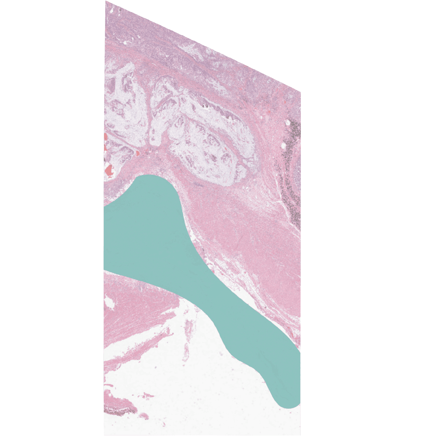
Colon cancer tissue classificaion

This AI model supports segmenting of various tissue types in colorectal cancer, including tumor regions, stroma, necrotic areas, nerves, fat, and muscle tissues. This advanced segmentation capability is invaluable in an end-to-end workflow, facilitating detailed cell counts and spatial analysis within tumor regions compared to surrounding tissues.

This AI model supports segmenting of various tissue types in colorectal cancer, including tumor regions, stroma, necrotic areas, nerves, fat, and muscle tissues. This advanced segmentation capability is invaluable in an end-to-end workflow, facilitating detailed cell counts and spatial analysis within tumor regions compared to surrounding tissues.

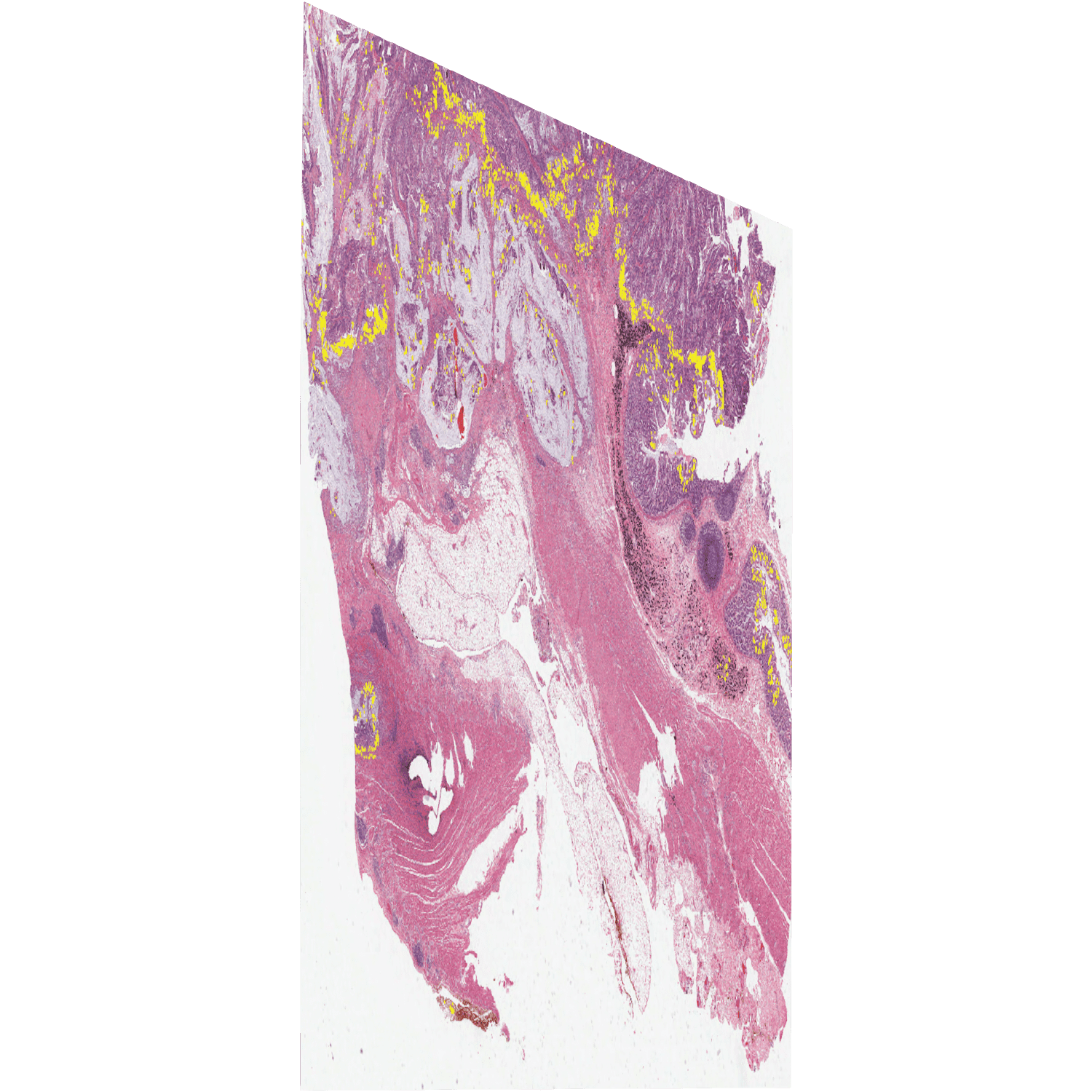
Nuclie segmentation

This AI model is capable of identifiing and segmenting different types of cells in whole-slide images. The model is tested in breast and colon cancer and is able to identify neoplastic, epithelial, connective and dead cells.

This AI model is capable of identifiing and segmenting different types of cells in whole-slide images. The model is tested in breast and colon cancer and is able to identify neoplastic, epithelial, connective and dead cells.

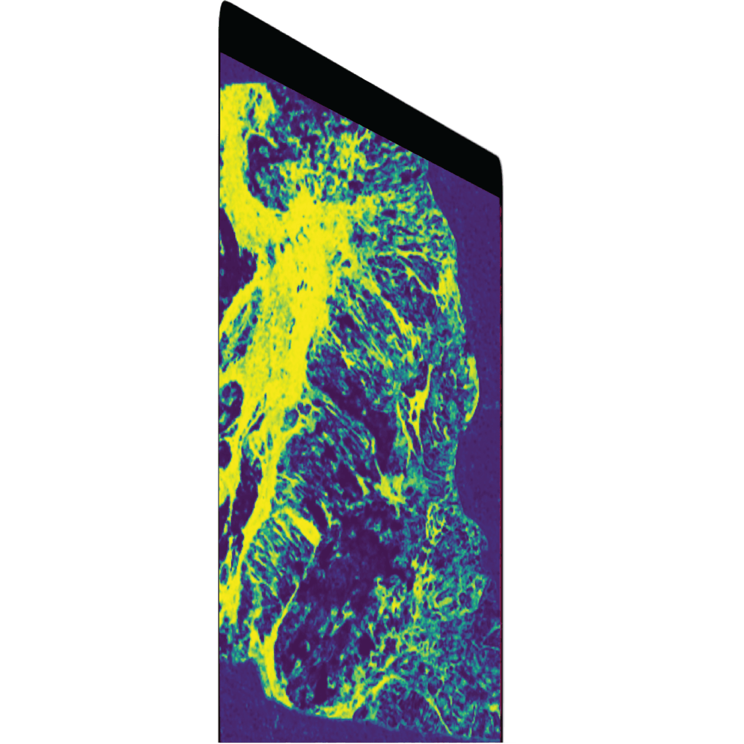
TME analysis

Orca Hub offers a set of pre-trained AI models to analyse the tumor microenvironment. This includes models for identifying and segmenting eosinophils, lymphocytes and plasma cells within tumor and stroma regions.

Orca Hub offers a set of pre-trained AI models to analyse the tumor microenvironment. This includes models for identifying and segmenting eosinophils, lymphocytes and plasma cells within tumor and stroma regions.

Deep Statistical and Spatial Analysis

Count, visualise and correlate with disease outcome

Perform large-scale analysis on the cell level to classify thousands of different cell types. Extract relevant statistics to support biomarker identification and correlation with clinical outcomes.

Spatially Analyse cells and tissues to understand the TME

Get a better understanding of the tumor microenvironment by analysing and quantifying cell-cell interactions and cell-tissue interactions at a large scale

Read the Paper

Explore, Orca™

With a powerfull AI engine optimised to unlock the full power of WSI analysis, ORCA accelerates workflows like never before. Now AI at your finguretips — from pretrained AI suite to your own custom models, all at the speed, accuracy and ease of use so you can focus on what matters the most.

Get in Touch

Learn more about Orca™. Schedule a call with our team.


contact@spatialx.ai

+44 (0) 7742 2138 81

1 Ariel Way

W12 7SL
London, United Kingdom

SpatialX

© 2024. All rights reserved

Keep in touch

Get in Touch

We're here to help. Schedule a call with our team.


contact@spatialx.ai

+44 (0) 7742 2138 81

1 Ariel Way

W12 7SL
London, United Kingdom

SpatialX

© 2024. All rights reserved

Keep in touch

Built by Pathologists,

for Pathologists.

We're here to help. Schedule a call with our team.


contact@spatialx.ai

+44 (0) 7742 2138 81

5A Clarendon Rd
W11 4JA
London, United Kingdom

SpatialX

© 2024. All rights reserved

Keep in touch

Built by Pathologists,

for Pathologists.

We're here to help. Schedule a call with our team.


contact@spatialx.ai

+44 (0) 7742 2138 81

5A Clarendon Rd
W11 4JA
London, United Kingdom

SpatialX

© 2024. All rights reserved

Keep in touch